Cardiac cycle


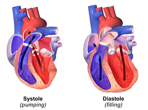
The cardiac cycle is the performance of the human heart from the beginning of one heartbeat to the beginning of the next. It consists of two periods: one during which the heart muscle relaxes and refills with blood, called diastole (die-ASS-toe-lee), followed by a period of robust contraction and pumping of blood, dubbed systole (SIS-toe-lee). After emptying, the heart immediately relaxes and expands to receive another influx of blood returning from the lungs and other systems of the body, before again contracting to pump blood to the lungs and those systems. A normally performing heart must be fully expanded before it can efficiently pump again. Assuming a healthy heart and a typical rate of 70 to 75 beats per minute, each cardiac cycle, or heartbeat, takes about 0.8 seconds to complete the cycle.[1]
Cardiac cycle
CG Heart.gif
OrganismsAnimalia*
Biological systemCirculatory system
HealthBeneficial
ActionInvoluntary
MethodBlood is allowed to enter relaxed ventricle chamber from vein through venous valve. Heart muscle contracts ventricle chamber and blood is expelled through arterial valve to artery.
OutcomeCirculation of blood
Frequency60–100 per minute (Humans)
Duration0.6–1 second (Humans)
*Animalia with the exception of PoriferaCnidariaCtenophoraPlatyhelminthesBryozoanAmphioxus.
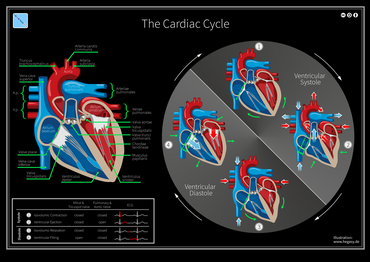
There are two atrial and two ventriclechambers of the heart; they are paired as the left heart and the right heart—that is, the left atrium with the left ventricle, the right atrium with the right ventricle—and they work in concert to repeat the cardiac cycle continuously, (see cycle diagram at right margin). At the "Start" of the cycle, during ventricular diastole–early, the heart relaxes and expands while receiving blood into both ventricles through both atria; then, near the end of ventricular diastole–late, the two atria begin to contract (atrial systole), and each atrium pumps blood into the ventricle 'below' it.[2] During ventricular systole the ventricles are contracting and vigorously pulsing (or ejecting) two separated blood supplies from the heart—one to the lungs and one to all other body organs and systems—while the two atria are relaxed (atrial diastole). This precise coordination ensures that blood is efficiently collected and circulated throughout the body.[3]

The cardiac cycle and Wiggers diagramEdit

The time-wise increases and decreases of the heart's blood volume (see Wiggers diagram), are also instructive to follow. The red-line tracing of "Ventricular volume" provides an excellent track of the two periods and four stages of one cardiac cycle. Starting with the Diastole period: the low-volume plateau of "Isovolumic relaxation" stage, followed by a rapid rise and two slower rises, all components of the "Inflow stage"—increasing to the high-volume plateau of the "Isovolumic contraction" stage; (find the label at left side of diagram). Then, the Systole, including the high "Isovolumic contraction" stage to the rapid decrease in blood volume (i.e., the vertical drop of the red-line tracing) which signifies the emptying of the ventricles during the "Ejection" stage of the completed cycle—all equal to one heartbeat.

StagesEdit

StageAV valves*Semilunar valvesStatus of ventricles and atria; and blood flow
1 Isovolumic relaxation
closed
closed
• semilunar (pulmonary and aortic) valves close at end of ejection stage; blood flow stops.
2a Inflow: (Ventricular filling)
open
closed
• ventricles and atria together relax and expand; blood flows to the heartduring ventricular and atrial diastole.
2b Inflow: (Ventricular filling with Atrial systole#)
open
closed
• ventricles relaxed and expanded; atrial contraction (systole) forces blood under pressure into ventricles during ventricular diastole–late.
3 Isovolumic contraction
closed
closed
• AV valves close at end of ventricular diastole; blood flow stops; ventricles begin to contract.
4 Ejection: Ventricular ejection
closed
open
• ventricles contract (ventricular systole); blood flows from the heart—to the lungs and to rest of body during ventricular ejection.

PhysiologyEdit

Heart electrical conduction systemEdit

In a healthy heart all activities and rests during each individual cardiac cycle, or heartbeat, are initiated and orchestrated by signals of the heart's electrical conduction system, which is the "wiring" of the heart that carries electrical impulses throughout the body of cardiomyocytes, the specialized muscle cells of the heart. These impulses ultimately stimulate heart muscle to contract and thereby to eject blood from the ventricles into the arteries and the cardiac circulatory system; and they provide a system of intricately-timed and persistent signaling that controls the rhythmic beating of the heart muscle cells, especially the complex impulse-generation and muscle contractions in the atrial chambers. The rhythmic sequence (or sinus rhythm) of this signaling across the heart is coordinated by two groups of specialized cells, the sinoatrial (SA) node, which is situated in the upper wall of the right atrium, and the atrioventricular (AV) node located in the lower wall of the right heartbetween the atrium and ventricle. The sinoatrial node, often known as the cardiac pacemaker, is the point of origin for producing a wave of electrical impulses that stimulates atrial contraction by creating an action potential across myocardium cells.[9][10]

Diastole and systole in the cardiac cycleEdit

Cardiac diastole is the period of the cardiac cycle when, after contraction, the heart relaxes and expands while refilling with blood returning from the circulatory system. Both atrioventricular (AV) valves open to facilitate the 'unpressurized' flow of blood directly through the atria into both ventricles, where it is collected for the next contraction. This period is best viewed at the middle of the Wiggers diagram—see the panel labeled "Diastole". Here it shows pressure levels in both atria and ventricles as near-zero during most of the diastole. (See gray and light-blue tracings labeled "Atrial pressure" and "Ventricular pressure"—Wiggers diagram.) Here also may be seen the red-line tracing of "Ventricular volume", showing increase in blood-volume from the low plateau of the "Isovolumic relaxation" stage to the maximum volume occurring in the "Atrial systole" sub-stage.

Atrial systoleEdit

Atrial systole is the contracting of cardiac muscle cells of both atria following electrical stimulation and conduction of electrical currents across the atrial chambers (see above, Physiology). While nominally a component of the heart's sequence of systolic contraction and ejection, atrial systole actually performs the vital role of completing the diastole, which is to finalize the filling of both ventricles with blood while they are relaxed and expanded for that purpose. Atrial systole overlaps the end of the diastole, occurring in the sub-period known as ventricular diastole–late (see cycle diagram). At this point, the atrial systole applies contraction pressure to 'topping-off' the blood volumes sent to both ventricles; this atrial kickcloses the diastole immediately before the heart again begins contracting and ejecting blood from the ventricles (ventricular systole) to the aorta and arteries.

Ventricular systoleEdit

©Atps.Plus™

Comments

Popular posts from this blog

Medical About

Open main menu ©Aah.Towfiqβ Search Edit Watch this page Read in another language Medicine This article is about the science of healing. For medicaments, see medication. For other uses, see Medicine (disambiguation). "Academic medicine" redirects here. For the journal, see Academic Medicine (journal). Medicine  is the science and practice of the diagnosis, treatment, and prevention of disease. Medicine encompasses a variety of health care practices evolved to maintain and restore health by the prevention and treatmentof illness. Contemporary medicine applies biomedical sciences, biomedical research, genetics, and medical technology to diagnose, treat, and prevent injury and disease, typically through pharmaceuticals or surgery, but also through therapies as diverse as psychotherapy, external splints and tr...

Welftion Application Form

click Now And Please LIKE COMMENT Facebook   Twitter   Instagram   Linkedia   WhatsApp   imo   Google Site   Facebook  Group   YouTube   Mesanger   Google  Search  Welftion Human Welfare  Association  Search     ওয়েলফশন মানবকল্যাণ সংঘ সার্চ ওয়েলফশন Loading… Loading… ওয়েলফশন তোমার আমার সংগঠন।  #Welftion #وعلفشن #ওয়েলফশন    " Stay connected with us"  আমাদের সাথে যুক্ত থাকুন   Twitter :- https://www.twitter.com/welftion  🚩Page : https://www.facebook.com/WELFTION  🔗 https://sites.google.com/view/welftion/  Websites Pest On :  HTML :-  <iframe src="https://docs.google.com/forms/d/e/1FAIpQLSccW2Mo7HQUDRLk9HRJxLyWvWvFhqNHkCseWv9-naFu1fXJcA/viewform?embedded=true" width="640" height="1508" frameborder="0" marginheight="0" marginwidth="0">Loading…</iframe> html °° Blog &  Website : <iframe src="https://docs.goo...

welftion

©Atps.©     #Welftion #ওয়েলফশন #وعلفشن || Welftion ওয়েলফশন   بِسْمِ اللّهِ الرَّحْمـَنِ الرَّحِيمِ  "সততায় আমরা" "কল্যাণে আমরা" "শান্তিতে আমরা"   ওয়েলফশন ~ মানবকল্যাণ সংঘ   বিশ্বের কল্যাণকামী মানুষের একটি সম্মিলিত সংগঠন।   প্রবর্তন : ০৪ ফেব্রুয়ারি ২০১৯ খ্রিঃ  E-mail : welftion@yahoo.com  ভূমিকাঃ সংগঠন একটি সামাজিক প্রক্রিয়া। যেখানে একদল মানুষ একটি সাংগাঠনিক কাঠামোর অন্তভুক্ত হয়ে নিদিষ্ট কিছু লক্ষ্য ও উদ্দেশ্য বাস্তবায়নে সর্বদা নিরন্তর। ওয়েলফশন - মানবকল্যাণ সংঘ এর ব্যাতিক্রম নয়। ওয়েলফশন একটি মানবসেবা ও মানব উন্নয়ন মূলক সামাজিক সংগঠন । যেখানে বিশ্বের কল্যাণ কামী দল & ব্যক্তি একত্রিত হয়ে। স্ব স্ব এলাকার শিক্ষা সচেতনতা, বেকারত্ব দূরীকরণ, আর্থ-সামাজিক উন্নয়ন,ক্রিয়া, সাংস্কৃতিক পরিবেশ, আত্ম-কর্মসংস্থান, মানবাধিকার এবং সারাবিশ্বে শান্তি প্রতিষ্ঠাসহ পারষ্পরিক সহযোগিতা, সহমর্মিতা ও ভ্রাতৃত্বের সেতু স্থাপন তথা আদর্শ সমাজ জীবন গড়ে তোলার দৃঢ় প্রত্যয়ে সামাজিক, উন্নয়নশীল ও উন্নয়নমূলক স্বেচ্ছাসেবী সংগঠন হিসেবে সততা ও কল্যাণের মাধ্যমে শান্তি প্রতিষ্ঠার ল...